

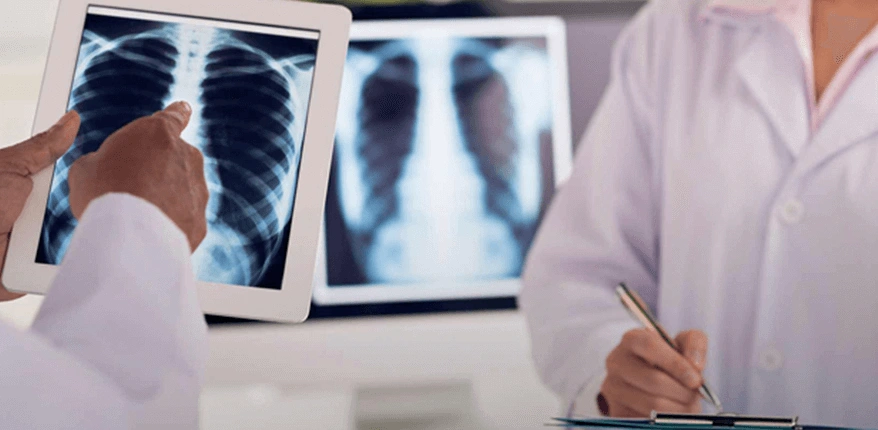
Radiology
Our hospital is equipped with the most advanced high-tech radiological devices, allowing the performance of all diagnostic and interventional radiological procedures. The Radiology Department has the technical infrastructure and experienced medical staff to meet all diagnostic and treatment-related requests from patients and physicians. The staff takes maximum care to ensure patient comfort during procedures.
Radiologists have the knowledge and experience not only to perform diagnostic procedures such as general radiology, computed tomography, mammography, panoramic X-ray, digital fluoroscopy, magnetic resonance imaging, ultrasonography, color Doppler ultrasonography, and angiography, but also to carry out therapeutic interventional procedures. Operating 24/7, the department provides immediate reporting to speed up the diagnostic and treatment process.
Ultrasonography (US) and Doppler Ultrasonography
All ultrasonographic examinations such as abdominal, pelvic, kidney, thyroid, thoracic, breast, hip, ocular, and cranial ultrasonography are performed. Transrectal US, transvaginal US, and intraoperative US are also among the services provided.
In addition, fine-needle aspiration and biopsies, fluid aspirations, and catheter drainage procedures are performed under ultrasound guidance. Vascular structures and blood flow can be evaluated in detail using color Doppler ultrasonography. These include upper and lower extremity arterial and venous system Doppler, portal Doppler, carotid and vertebral artery Doppler, orbital and scrotal Doppler, gynecological and obstetric Doppler, renal artery Doppler, and graft evaluations.
All examinations are performed by highly experienced physicians. Patient satisfaction is a primary principle of the department; patients can receive same-day appointments, and waiting times are kept as short as possible.
Double-contrast and biphasic gastrointestinal system studies, as well as enteroclysis examinations, can be performed. Genitourinary system studies include excretory urography, cystography, voiding cystourethrography, and retrograde urography. All hysterosalpingographies are performed in the presence of an obstetrician and interpreted by radiologists. In fluoroscopic studies, the focus is on achieving the highest quality images with minimal radiation exposure.
Mammography
Mammography is a special X-ray examination used for the diagnosis of breast cancer and other benign breast diseases. Mammography services include screening of asymptomatic patients, diagnostic evaluation of women with symptomatic breast disease, stereotactic preoperative breast localization, and consultations.
The technological features of the devices allow optimal magnification and compression techniques with minimal radiation exposure. In addition to screening and diagnostic mammography, galactography, ultrasound-guided breast localization, fine-needle aspiration, and biopsies are successfully performed.
Images from all examinations are transferred to the PACS (Picture Archiving and Communication System) for evaluation and storage by radiologists. Integration with the hospital information system (HIS) enables rapid access to results from all outpatient clinics and wards, as well as easy consultation when necessary.
Computed Tomography (CT)
CT provides detailed cross-sectional images of any part of the body and is used to diagnose many conditions, from soft tissue diseases to fractures. It is frequently preferred when other imaging methods such as direct radiography and ultrasonography fail to provide sufficient information. Spiral or sequential scans of the brain, spine, thorax, abdomen, bones, and pelvis can be performed. CT-guided biopsies and interventional procedures are also performed to reduce the need for surgery and obtain tissue diagnosis of lesions.
Magnetic Resonance Imaging (MRI)
All neurological studies (brain, neck, and spinal column), vascular angiography (peripheral, intracranial, carotid, and aortic), abdominal, thoracic, and cardiac examinations, breast MRI, musculoskeletal system examinations, MR cholangiopancreatography (MRCP), fetal MRI, and functional MRI are performed.
Functional MRI techniques include blood oxygen level-dependent imaging (BOLD), diffusion and perfusion MRI, MR spectroscopy, and cerebrospinal fluid flow analysis. Breast MRI is performed in cases where mammography and ultrasound cannot provide a definitive diagnosis of breast cancer. MRCP provides non-invasive imaging of the biliary and pancreatic ducts and is increasingly replacing diagnostic ERCP examinations. MRI procedures requiring anesthesia for patients of all ages are performed under the supervision of an anesthesiologist.
Whole-Body MRI with 3 Tesla
What is 3 Tesla Whole-Body MRI?
3 Tesla whole-body MRI is an imaging method used to examine the entire body in detail from head to toe.
What is it used for?
Whole-body MRI can be used to diagnose, monitor, or assess a variety of health conditions, including cancer, heart diseases, brain disorders, spinal problems, and liver and kidney diseases. It can be used for diagnostic purposes in healthy individuals as well as in the follow-up of cancer patients.
How is it performed and how long does it take?
Whole-body scanning is performed using a 3 Tesla MRI device. The patient lies on a table that moves into a long tunnel, and the scanner images different parts of the body using a magnetic field and radio waves. The scan takes approximately 45–50 minutes.
What makes the whole-body MRI program different?
- 3 Tesla MRI provides a holistic view of the entire body.
- Unlike imaging methods that focus on a single organ, whole-body MRI can evaluate changes in multiple organs and systems in one scan.
- It does not involve radiation exposure, making it suitable for patients wishing to avoid radiation, especially children.
- It is non-invasive and requires no surgical intervention, unlike certain other procedures such as biopsies.
Frequently Asked Questions
What preparation is needed before a whole-body MRI?
Whole-body MRI generally does not require the use of contrast material. However, contrast injection may be necessary in specific cases. It is important to remain still during the scan, as movement can affect image quality. Metal objects should be removed before entering the scan area. Patients should inform their physician if they are pregnant, have a pacemaker, or use other medical devices, so the scan can be planned accordingly.
Does whole-body MRI cause pain?
No, it does not cause pain. Only the noise of the device is heard, for which ear protection is provided. It is a non-invasive imaging method with no surgical intervention.
For which patients is whole-body MRI recommended?
- Cancer: To detect, evaluate spread, or assess metastases, including early-stage detection for timely treatment.
- Lymphoma: To evaluate enlargement, involvement, or metastasis in lymph nodes throughout the body.
- Rheumatologic Diseases: To assess joint damage, inflammation, or other changes in conditions affecting joints, muscles, and connective tissue.
- Inflammatory Diseases: Such as Crohn’s disease, multiple sclerosis, and sarcoidosis.
- Metabolic Diseases: To monitor or diagnose rare metabolic disorders such as Wilson’s disease or Gaucher disease.
Does whole-body MRI involve radiation?
No. MRI uses magnetic fields and radio waves and does not emit harmful radiation.
Is a chemical injection required before the scan?
Generally, no. In certain cases, contrast material may be used to better visualize specific structures or pathologies.